Periodontal Ehlers-Danlos Syndrome (pEDS)
The Ehlers-Danlos syndromes (EDS) are a group of 13 related conditions that affect connective tissue (1). Connective tissue is found everywhere in the body. It connects other types of tissues, separates them, and supports them. Examples of connective tissue include bone, cartilage, fat, blood, and lymphatic tissue. When connective tissue is abnormal because of a disease, a variety of problems result.
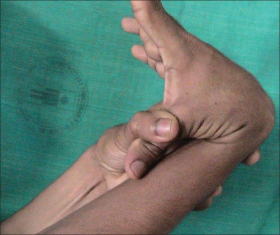
For example, certain problems happen in all of forms of EDS. The first is joint hypermobility (often called loose or double joints). People with EDS can often move their joints in ways that are impossible for most unaffected people. The photograph at the bottom of this page has an example (note that many people without EDS have loose joints, so having joint hypermobility does not mean that you have EDS). The second thing that happens in every type of EDS is stretchy (hyperextensible) skin. This feature is nearly universal in some types of EDS (such as type 1, the classical form). In our literature survey, we found that stretchy skin occured in less than half of people with the hypermobile and vascular forms, more than half of patients with the periodontitis type, and in a large majority of people with the arthrochalasia type (>80%).
Many forms of EDS happen because of mutations in genes that make collagen, which can be thought of as both holding the body together and strengthening it. Collagen gives structure to muscles, tendons, skin and bones. In most people, it is abundant in these tissues. In people with EDS, collagen may not be as strong as it should be, or there may not be enough of it. For example, skin and joints may be loose because they don't have enough collagen to hold them together securely, or because the collagen they have is weaker than it should be.
Doctors and other medical professionals assess joint hypermobility with a scoring system called the Beighton score, which was developed in the early 1970s (2). This simple system scores your ability to move a joint past certain angles. The highest score is 9 points, and higher scores mean greater joint hypermobility. Generally speaking, scores of 4 or higher (at any time, past or present) indicate joint hypermobility. Many EDS patients have scores of 8 or 9, although many have lower scores, and a minority do not have hypermobile joints. Interested readers may wish to see a basic description of the scale with a video and photos or a more technical description.
Joint hypermobility puts EDS patients at high risk for joint dislocations and partial dislocations (called subluxations). These problems can be painful, and in some cases, debilitating. Surgical procedures have been used to improve skeletal stability and improve quality of life; for examples and reviews, see references 3-5. However, experts tend to agree that non-surgical options should always be exhausted first, and careful discussions with doctors are important in this decision.
Tissue fragility is another common feature of EDS. This problem is very serious in the vascular type of EDS (vEDS). The type of collagen affected in vEDS plays a role in supporting blood vessels. When the integrity of their support system is affected, blood vessels can rupture spontaneously. This problem is especially dangerous because it affects medium and large blood vessels, as well as small ones. It also affects the bowel. Surgery on a person with vEDS must be performed with great care in order to avoid serious complications. This problem may also happen in kyphoscoliotic EDS (kEDS), but it is less common than in vEDS. Alternatively, while people with the hypermobile form (hEDS) are also prone to blood vessel ruptures, ruptures tend to occur in the small blood vessels. Tissue fragility can also lead to hernias and problems in pregnancy. Fragile skin is also a common manifestation of EDS, especially in people with the classic, kyphoscoliotic, and periodontitis types.
Tissue fragility in EDS can also lead to hernias and problems in pregnancy. Fragile skin is also a common manifestation of EDS, especially in people with the classic, kyphoscoliotic, and periodontitis types.
A new classification system for EDS was released in 2017 (1). It's called the 2017 International Classfication for the Ehlers-Danlos Syndromes. This new system names 13 different subtypes of EDS. It relies primarily on descriptive terms for different types of EDS (e.g. vascular EDS, meaning that it affects blood vessels prominently). Two other systems existed before this one. They were named Villefanche [1998] and Berlin [1988]. Both systems used names and numbers. The numbers in the new system are different from the old ones, and we've included the old ones here. Advances in genetics have allowed to researchers to find genes that cause different subtypes of EDS, which has allowed them to separate the old classic subtype (numbered 1 & 2) of EDS into 2 different subtypes.
- 1. Classical EDS (cEDS; formerly type 1/2)
- 2. Classical-like EDS (cEDS; formerly type 1/2)
- 3. Cardiac-valvular (cvEDS)
- 4. Vascular (vEDS)
- 5. Hypermobile (hEDS; formerly type 3)
- 6. Arthrochalasia (aEDS; formerly type 7a/b)
- 7. Dermatosparaxis (dEDS; formerly type 7c)
- 8. Kyphoscoliotic (kEDS; formerly type 6)
- 9. Brittle Cornea syndrome (BCS)
- 10. Spondylodysplastic (spEDS)
- 11. Musculocontractural (mcEDS)1
- 12. Myopathic (mEDS); type 12
- 13. Periodontal (pEDS; formerly type 8)
EDS affects women more commonly than men. In the literature survey we did for our analytics tool, we counted the number of male and female patients while we were characterizing in six different types of EDS. Altogether, we analyzed data from ~1,200 patients. Sex information was available for ~1,100. The table below shows that although more EDS patients are women in each type of EDS, the female:male ratio varied between each type. For example, females dominated the individuals reported with the hypermobility type, while males comprised a substantial minority of kyphoscoliotic and arthrochalasia patients. The distribution was roughly even in the periodontal type. However, the low number of case reports for these three types of EDS may have affected the outcome of this small analysis.
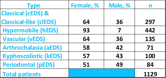
Above: Percentages of male and female patients with different types of EDS. Source: FDRF via published literature.
According to NIH's Genetics Home Reference (see link at right), the prevalence of all forms of EDS is roughly 1 in 5,000 worldwide. Type 8 is very rare, and its prevalence is not known.
EDS is a generally characterized by hypermobile joints and skin. However, these two clinical features are not uniformly prevalent in type 8 and are generally not as severe as they are in other types of EDS. There is also considerable variation in joint hypermobility, with some patients having no hypermobility or hypermobility in a limited number of joints.
Clinical information
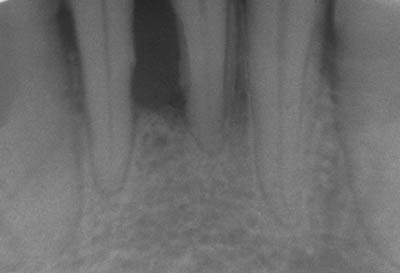
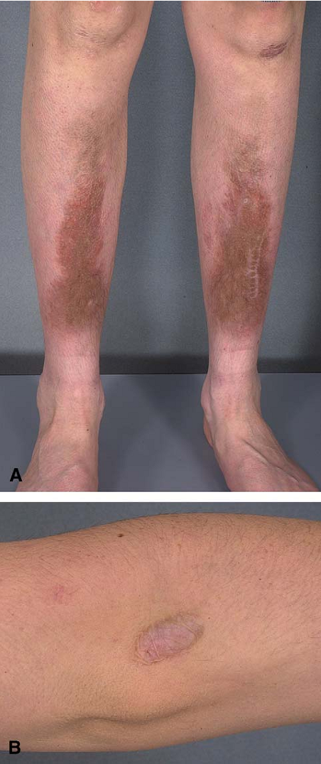
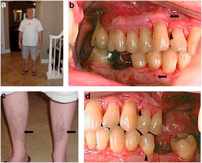
The most striking features of pEDS are dental and dermatological. Patients tend to have severe periodontal disease and fragile, easily bruised skin, especially on the lower legs. The presence of cigarette paper scars and bruised-looking tibial lesions (called pretibial plaques) are very common in peope with pEDS. The photos on this page illustrate these problems.
As with all other forms of EDS, the clinical features of pEDS overlap with other forms of EDS. In particular, there is some overlap between pEDS and the vascular type of EDS. The lack of fragile blood vessels may help distinguish the two (see Differential Diagnosis, below). In general, the following features occur commonly in this form of EDS:
- Periodontal disease: - Gum recession (see photos)
- Easy bruising
- Brownish-yellowish shin lesions (pretibial plaques; see photo)
- Fragile skin (lower legs especially; may split with minimal trauma)
- Flat feet, especially when standing
- Smooth, velvety skin
- Loose or stretchy skin (roughly half of patients)
- Joint hypermobility (may not be extreme; may be limited to the hands)
- Arachnodactyly (long, slender fingers)
- A Marfanoid habitus (tall and slender, with arachnodactyly; armspan may be greater than height)
- Hernia(s)
Signs and symptoms of pEDS
- Alveolar bone loss (loss of bone that forms tooth sockets; see photos)
- Loss of permanent teeth (often begins in adolescence; sometimes sooner)
- Inflamed, swollen gums
- Loss of some or all permanent teeth, often at a young age
- Many cavities
We found reports of cardiac abnormalities as well. Mitral valve prolapse was the most common reported problem.
Some patients with pEDS have distinctive facial features. These features are not as obvious, common, or numerous as they are in patients with other forms of EDS, such as vascular EDS. However, awareness of the constellation of facial features associated with each type may help with diagnosis. In EDS VIII, they include the following:
- A triangular face
- Prominent eyes
- A distinctive nose (long, with narrow root)
- Some patients may show premature aging
Facial features that may occur in pEDS
Diagnosis and Testing
pEDS is an autosomal dominant disorder. This term means that it is usually passed from one affected parent to a child. However, it can also occur sporadically in a person without an affected parent. In our analysis, information on the family history of EDS or EDS-like connective tissue disease was available for 70 patients. Sixty, or 86%, had an affected parent. There was no history of an EDS-like disorder in the families in the remaining 10 people.
The criteria below were established as part of the 2017 classification system (1). For a diagnosis, a patient must have the first major criteria OR the second one AND at least two other major criteria and one minor criterion. Identification of a mutation is also essential to verify a diagnosis based on the clincal criteria.
Major criteria
- Severe and intractable periodontitis with onset in childhood or adolescence
- Lack of attached gingiva (loose gums)
- Pretibial plaques
- Family history of the disease in a first-degree relative (parents, siblings, children)
Minor criteria
- Easy bruising
- Joint hypermobility, mostly distal joints (joints far from the center of the body, like hands/feet)
- Skin hyperextensibility and fragilityk, abnormal scarring (wide or atrophic)
- Increased number of infections
- Hernias
- Marfanoid facial features
- Premature aging appearance (acrogeria)
- Prominent blood vessels (vasculature)
Mutations in the genes C1R and C1S are associated with pEDS (reviewed in reference 1). Previous work in a large study in Sweden had implicated a region of chromosome 12 (12p13) with pEDS (10), which is where these genes are located. Another research group found this connection in a patient from the United States and and a connection between pEDS and chromosome 9 in other US patients (7,8). Because of the lack of gene-disease associations, the diagnosis of pEDS is primarily clinical, with laboratory tests for collagen being useful for ruling out other forms of EDS.
pEDS is generally suspected in patients with periodontal disease, shin lesions such as those pictured here, and fragile, easily bruised skin. In addition, hypermobile joints and stretchy skin occur in pEDS. While these clinical features are less common in pEDS than in most other forms of EDS, their presence, along with periodontal disease and shin lesions is suggestive of pEDS (but not definitive; 8). In our survey of the literature, roughly 75% of pEDS patients had lax joints and slightly more than half had stretchy skin.
It is important to remember that there is a lot of overlap between the different forms of EDS. As noted above, there is overlap between pEDS and the vascular type of EDS (EDS type 4). In particular, periodontal disease has been reported in patients with vascular EDS (see Differential Diagnosis, below; 9). This fact means that, in the absence of laboratory testing, definitive subcategorization of EDS in a patient may be difficult or even impossible.
Differential Diagnosis
The clinical features of the vascular form of EDS overlap with pEDS. Specifically, a number of clinical features are seen in both:
- Hyperextensible skin
- Fragile skin
- Thin/transluecent skin
- Easy bruising
- Joint hypermobility
- Flat feet
- Periodontal disease
- Blood vessel fragility
While most items on the above list are relatively common in both types, the prevalence of periodontal disease is low in the vascular form, and one study noted that it is not a good diagnostic indicator for EDS-vascular (9). In addition, the prevalence of blood vessel fragility (apart from fragile capillaries associated with easy bruising) appears to be low in pEDS. In our analysis of published cases, this problem was mentioned in only one patient out of 20 in which the condition was discussed (6). Thus, these two characteristics may help distinguish the two conditions. In addition, laboratory tests are available for EDS-vascular, which is associated with mutations in the gene COL3A1. Thus, gene sequencing can either make a definitive diagnosis of EDS-vascular or rule it out. EDS-vascular patients are prone to spontaneous ruptures of large blood vessels that may be fatal. For this reason, clinicians may wish to use genetic testing to definitively rule it out in any patient suspected of having EDS.
pEDS may be distinguished from the kyphoscoliotic type by genetic testing: the kyphoscoliotic form is caused by mutations in PLOD1. In addition, people with the kyphoscoliotic form tend to have severe spinal deformities. These deformities occur in pEDS, but are not generally so severe as they are in the kyphoscoliotic type.
The classical and classical-like forms of EDS range in severity from mild to relatively severe. Like many pEDS patients, cEDS patients have hypermobile joints and stretchy skin. Some features may help distinguish the two disorders. Most importantly, cEDS patients tend to have severe skin involvement that can be disfiguring. Skin involvement in pEDS patients can be severe on the legs, but extreme skin fragility and the lesions commonly seen on the shins of pEDS patients do not appear to extend to other parts of the body. Molluscoid pseudotumors or subcutaneous spheroids (see pictures on the classic form page) have not been reported in pEDS patients, but occur in more than hlf of EDS-classic patients.
The hypermobile form of EDS (hEDS) is a relatively mild form of EDS. As its name implies, joint hypermobility is the primary feature of this form of EDS. Unlike pEDS, cigaratte paper scars do not appear to be a general feature of hEDS. In our analysis, they were cited in more than 75% of pEDS patients.
People with the arthrochalasia type of EDS suffer frequent dislocations, and many are born with hip dislocations. This feature can help distinguish this condition from pEDS.
Overall, joint hypermobility occurs in a variety of medical conditions, and many people without a medical condition have lax joints. In fact, when not part of a disorder, hypermobile joints confer an advantage in sports such as gymnastics and ice skating, as well as in certain forms of dance.
Marfan syndrome. Most medical conditions involvong lax joints are relatively easy to distinguish from EDS, but some are not. Marfan syndrome is an example. Like EDS, Marfan syndrome is a connective tissue disorder and patients have lax joints. Marfan patients generally have a body type called a Marfanoid body habitus. This term means that a person is tall and slender, with long arms, hands, fingers, legs, feet and toes. A Marfanoid body habitus occurs in a minority of EDS-H patients, but it does not appear to be generally associated with classic EDS. The Marfan Foundation has many photographs of Marfan patients of different ethnicities. Unlike EDS patients, Marfan patients do not appear to have fragile skin and blood vessels.
Cutis laxa. Superficially, the stretchy skin found in most forms of EDS may be confused with cutis laxa type 1 and type 2, as stretchy skin is also a feature of these conditions. However, the skin in cutis laxa patients tends to sag and does not return to its normal position after being extended. Cutis Laxa patients often have a prematurely aged appearance.
References
- 1. Malfait F et al. (2017) The 2017 International Classification of the Ehlers-Danlos Syndromes Am J Med Genet C 175(1):8-26. Abstract on PubMed.
- 2. Beighton P et al. (1973) Articular mobility in an African population. Ann Rheum Dis 32(5):413-418. Full text on PubMed.
- 3. Cole AS et al. (2000) Recurrent instability of the elbow in the Ehlers-Danlos syndrome. A report of three cases and a new technique of surgical stabilisation. J Bone Joint Surg Br 82(5):702-704. Full text from publisher.
- 4. Weinberg J et al. (1999) Joint surgery in Ehlers-Danlos patients: results of a survey. Am J Orthop 28(7):406-409. Abstract on PubMed.
- 5. Shirley ED et al. (2012) Ehlers-Danlos syndrome in orthopaedics. Etiology, diagnosis, and treatment implications. Sports Health 4(5):394-403. Full text on PubMed.
- 6. Rahman N et al. (2003) Ehlers-Danlos syndrome with severe early-onset periodontal disease (EDS-VIII) is a distinct, heterogeneous disorder with one predisposition gene at chromosome 12p13. Am J Hum Genet 73198-204. Full text on PubMed.
- 7. Reinstein E et al. (2011) Ehlers-Danlos type VIII, periodontitis-type: further delineation of the syndrome in a four-generation pedigree. Am J Med Genet Part A 155:742-747. Abstract on PubMed.
- 8. Reinstein E et al. (2013) Ehlers-Danlos syndrome type VIII is clinically heterogeneous disorder associated primarily with periodontal disease, and variable connective tissue features. Eur J Hum Gen 21:233-236. Full text on PubMed.
- 9. Ferré FC et al. (2012) Oral phenotype and scoring of vascular Ehlers-Danlos syndrome: a case-control study. BMJ Open 2(2):e000705. doi: 10.1136/bmjopen-2011-00070. Full text on PubMed.
- 10. Cikla U et al. (2014) Fatal ruptured blood blister-like aneurysm of middle cerebral artery associated with Ehlers-Danlos yndrome type VIII (periodontitis type). J Neurol Surg Rep 75(2):e210-e213. Full text on PubMed.
- 11. Moore MM et al. (2006) Ehlers-Danlos syndrome type VIII: periodontitis, easy bruising, Marfanoid habitus, and distinctive facies. J Am Acad Dermatol 55(2 Suppl):S41-S45. Abstract on PubMed.
- 12. Ronceray S et al. (2013) Ehlers-Danlos syndrome type VIII: a rare cause of leg ulcers in young patients. Case Rep Dermatol Volume 2013:Article ID 469505. Full text on PubMed.
- 13. Bhat RM et al. (2014) Type VIII - Ehlers Danlos syndrome with café-au-lait macules: a rare variant. Indian J Dermatol 59(3):317. Full text on PubMed.
- 14. Bernard B (2005) Image found on the Wikimedia Commons.
