Friedreich Ataxia (FRDA)
Friedreich Ataxia, or FRDA, is a member of a group of disorders called hereditary ataxias. Hereditary ataxias are are characterized by gait abnormalities. Other problems include hand and speech clumsiness, and abnormal, jerky eye movements. All of these problems derive from abnormalities in the central and/or peripheral nervous systems. Specifically, there are often problems in the cerebellum, the spinal cord, and/or the peripheral nerves. These diseases are progressive, which means that they get worse with time.
There are many hereditary ataxias; FRDA is the most common. Others include ataxia-telangiectasia (A-T), which is the second most common member of the group. Other diseases in the group are categorized in subgroups, such as the spinocerebellar ataxias and the autosomal dominant cerebellar ataxias. Each subgroup has many members. For example, there are so many spinocerebellar ataxias, they are named with numbers (e.g. spinocerebellar ataxia type 1, spinocerebellar ataxia type 17, etc.).Other conditions are named for their clinical features (e.g. cerebellar ataxia, deafness, and narcolepsy, autosomal dominant).
As noted, FRDA is the most common hereditary ataxia. Overall estimates for its prevalence vary, with older estimates being 1 person in 50,000, and newer estimates being up to 1 person in 29,000 (1; reviewed in 2). According to a recent estimate that used the newer figures, there are roughly 9,000 people with FRDA in the United States (3).
FRDA has been documented in people of European, Middle Eastern, Indian, and North African descent. It does not appear to occur in native Americans, sub-Saharan Africans, and east Asians (although other forms of ataxia do occur in these groups; 4).
Clinical information
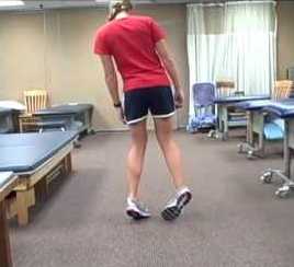
The primary feature of FRDA is progressive ataxia. Gait ataxia is also its most common presenting feature; patients develop an uncoordinated walk and may experience balance problems. See the photo at the right and the video link below for examples of gait ataxia. Other presenting features are listed below in decreasing order of frequency as a presenting feature (data combined from references 5 & 6):
- Gait ataxia (137/192 patients)
- Clumsiness (36/192)
- Weakness (lower limbs) (7/192)
- Tendency to trip or unsteady stance (6/192)
- Scoliosis (6/192)
- Cardiac problems (3/192)
- Tremor (2/192)
- Dysarthria (2/192)
- Vertigo (1/192)
The first signs of FRDA generally appear between the ages of 8 and 16 (5,6), although disease onset has been recorded as early as the toddler years and as late as the 70s. In general, earlier onset is associated with quicker progression of disease and later onset is associated with slower progression. However, this rule is not absolute, as there is great variability in FRDA, even among siblings.
The most common signs and symptoms of Friedreich ataxia are as follows. Note that FRDA patients do not have a common facial appearance.
- Ataxia (truncal and limb types)
- Absent or reduced tendon reflexes
- Extensor plantar reflexes (Babinski's sign)
- Peripheral neuropathy
- Atrophy of the cerebellum
- Nystagmus
- Speech disorder, most notably dysarthria
- Impaired joint position sense (patient does not know the position of a joint when he cannot see it)
- Impaired vibration sense (patient cannot feel a vibrating object)
- Scoliosis
- High arches (pes cavus); may get worse with time
- Weakness and muscle atrophy
- Cardiomyopathy
- Peripheral cyanosis
- Diabetes or glucose intolerance
The prognosis for FRDA patients has improved considerably in recent decades because of advances in therapies for cardiac problems and diabetes. Although no cure is available at this time, some patients live until their 60s or 70s. Physical therapy and rehabilitation programs can help maximize motor function, and surgery can help severe scoliosis and severe cardiac problems.
Diagnosis and Testing
FRDA is an autosomal recessive disorder caused by mutations in the gene frataxin (FXN). The term autosomal recessive means that the disorder is passed on when both parents contribute a copy of the mutated gene to their child. The link at the right provides information about labs that test for mutations in FXN.
At this time, FRDA is suspected based on clinical features and confirmed via testing for mutations in FXN. Clinically, there are two general types of FRDA typical and atypical). Typical FRDA accounts for roughly 75% of all cases, while several subtypes of atypical FRDA make up the rest. The subtypes are called late- and very-late-onset-FRDA, FRDA with retained reflexes, and FRDA in Acadians. The Acadian subtype is caused by a founder mutation in Canadians of French ancestry. Overall, the onset of disease is later in patients with atypical forms of FRDA and disease progression is slower. Both the typical and atypical forms are caused by mutations in FXN, and it is difficult to make predictions about disease outcomes based on genotype, as various other individual factors appear to affect the course of disease (7).
Clinicians diagnose FRDA by using a set of medical problems that are requirements for a diagnosis (5,8). For typical cases of FRDA, diagnosis requires that the following occur within 5 years of symptom onset:
- The patient is less than age 25 at symptom onset
- Progressive ataxia (limbs and gait) is present
- Knee and ankle jerks are absent or reduced
- Plantar responses are extensor (Babinski's sign)
- Motor nerve conduction velocity is >40 m/s, with reduced or absent sensory nerve action potential
In addition, scoliosis, absent/reduced reflexes in the upper limbs, an abnormal electrocardiogram, and loss of joint position and vibration sense are common problems that occur in >2/3 of cases. It is thought that the heart is affected (clinically or subclinically) in the vast majority of patients (8). Heart problems tend to get worse with time, and cardiomypoathy is the leading cause of death in FRDA (2). As a result, regular ECGs are recommended in FRDA patients (8).
In our survey we found that roughly 25% of FRDA patients had either diabetes or glucose intolerance. This finding agrees with those of others (8). Because complications of diabetes can have serious effects on health, it is important to monitor FRDA patients regularly for abnormalities of glucose metabolism. Diabetes may present in the usual ways (increased urination, increased thirst), and although patients may initially respond to oral drug therapy, they generally require insulin eventually (8).
Differential Diagnosis
There are several conditions in the differential diagnosis for FRDA. The most important of them is ataxia with vitamin E (α-tocopherol) deficiency, a treatable condition with symptoms that are very similar to FRDA's.
Ataxia with vitamin E deficiency (AVED). AVED is a hereditary condition that affects a person's ability to use vitamin E. Its clinical features are nearly idential to Friedreich ataxia: they include progressive ataxia, hand clumsiness, loss of joint position and vibration sense, and loss or reduction of tendon reflexes (9). Also like FRDA patients, people with AVED may have extensor plantar reflexes, a head tremor (titubation). Cardiomyopathy also occurs in AVED, but is less common than it is in FRDA. However, unlike FRDA patients, AVED patients have low levels of vitamin E in their blood. Reduced vitamin E is not a feature of FRDA. In addition, AVED occurs in east Asians, while FRDA does not. Muscle weakness and wasting are also not features of AVED (10).
AVED can be treated with vitamin E supplements (800-1200 mg per day; 10). Improvement of some symptoms occurs after treatment is begun, and no new problems appear during treatment (10). In addition, if treatment is begun in asymptomatic affected siblings of AVED patients, the siblings may not develop symptoms. Because AVED is a treatable condition and because it is so similar to FRDA, it is critical to test for it in any patient suspected of having FRDA. An analysis of serum vitamin E concentrations (low in AVED, normal in FRDA) and lipoprotein profiles (normal in AVED; abnormal in other ataxias) can help distinguish the two disorders. In addition, genetic sequencing is definitive, as AVED is caused by mutations in a gene called TTPA.
Charcot-Marie-Tooth disease (CMT). Charcot-Marie-Tooth neuropathy types 1 and 2 (CMT1 and CMT2) also resemble FRDA. Medical problems associated with CMT1 include distal muscle weakness and wasting/atrophy, sensory loss, and slow nerve conduction velocity. Like Friedreich ataxia, CMT1 patients tend to have high arches and walk with a foot drop. Symptom onset is usually between ages 5 and 25 years. Unlike FRDA, the vast majority of CMT1 patients do not eventually require wheelchairs, and life expectancy is not affected. Some CMT patients are clumsy and lose deep tendon reflexes as children. However, persistent dysarthria does not appear to be a feature of CMT1 or CMT2, although it has been reported in a few patients with CMTX, a third form of Charcot-Marie-Tooth disease. Extensor plantar reflexes do not appear to be a feature of CMT1 or CMT2, yet are very common in FRDA. Note that dysarthria and extensor plantar reflexes may develop over time in FRDA, and distinguishing it from CMT may be difficult as a result.
Unfortunately, there are may subtypes of CMT1 and 2, with more than 20 different genes associated with them. However, sequencing for FXN and the CMT genes can rule them in or out. In addition, most types of CMT1 and 2 are autosomal dominant, meaning that the presence of an affected parent is suggestive of CMT over FRDA. However, a relatively high percentage of the population carries a mutation in FXN, and cases of pseudo-dominant inheritance have occured when an FRDA patient has a child, and the other parent is a carrier (11). Thus, although an affected parent is suggestive that FRDA is not the diagnosis, it does not rule out FRDA.
Spinocerebellar ataxia with axonal neuropathy (SCAN1). SCAN1 is another disorder with many similarities to Friedreich ataxia. Symptom onset is usually in late childhood. Like FRDA patients, SCAN1 patients present with ataxia, which is followed by loss of tendon reflexes and peripheral neuropathy. Nystagmus, dysarthria, loss of vibration sense (hands and lower thigh), and high arches also develop. However, SCAN1 patients do not have cardiomyopathy or cerebellar atrophy. This disorder can be definitively distinguished from FRDA by gene sequencing for mutations in the gene TDP1.
Ataxia with oculomotor apraxia type 1 (AOA1). Like FRDA, AOA1 is a progressive neurological disorder involving ataxia. Most people with AOA1 begin to experience ataxia between the ages of 2 and 10 (average: ~4; 12) Ataxia is typically followed by oculomotor apraxia. This term means that patients have trouble moving their eyes horizontally in order to watch a moving object. They may have trouble with commencing eye motion, and may have to turn their heads in order to watch the object. This problem does not occur in Friedreich ataxia.
MRI of the cerebellum can also distinguish AOA1 and FRDA. In AOA1, cerebellar atrophy is visible in all patients (12). In FRDA, it is not.
Like FRDA patients, AOA1 patients also lose their tendon reflexes (e.g. knee jerk reflexes). They also develop peripheral neuropathy and choreiform movements jerky involuntary limb movements). We found reports of choreiform movements in FRDA patients during our literature survey, though they were reported only in a very small number of patients. AOA1 and FRDA patients also experience peripheral neuropathy, which may be severe, resulting in impaired mobility, atrophy and limb deformities. Another factor that can help distinguish AOA1 from FRDA is cognitive impairment, which is found in many AOA1 patients. This problem is not a feature of FRDA, although it does occur as would be expected in the poplation at large. In addition, eye blinking in AOA1 patients is exaggerated (12). As in FRDA, the clinical features of AOA1 vary from patient to patient. However, abnormalities of eye movement and the cerebellum on MRI can help distinguish it from FRDA. In addition, plantar reflexes are flexor in AOA1, while they are extensor in most FRDA patients (if the bottom of the foot is stroked, the toes curl inward toward the sole of the foot in AOA1 and flex backward above the foot in FRDA; inward is normal). Finding mutations in the gene APTX can also provide a definitive diagnosis of AOA1.
Abetalipoproteinemia (ABL). ABL, also called Bassen-Kornzweig syndrome, affects the body's ability to use fats that have been eaten. This is because people with ABL cannot transport fats out of their intestines. Like FRDA, ABL is an autosomal recessive disorder and is very rare. Prevalence is estimated at less than one person per million (13). People with ABL have very low levels of total and LDL cholesterols, and very low levels of triglycerides. These molecules are necessary for the absorption of vitamins A, E, and K from the intestines, and the lab tests show low levels of these vitamins. Vitamin E deficiency causes neurological signs that are similar to those of Friedreich ataxia and AVED (14). However, unliked AVED and FRDA, neurological signs are not the first problems to appear, a fact that can help distinguish ABL from them. The first clinical signs of ABL are generally gastrointestinal, and include steatorrhea (fatty stools), diarrhea with steatorrhea, and liver disease. AVED and FRDA do not generally cause these problems. In addition, laboratory studies of serum cholesterol (total and LDL) are normal in AVED/FRDA but abnormal in ABL. Serum vitamin E may be reduced in both AVED and ABL; levels are normal in FRDA.
References
- 1. Cossée M et al. (1997) Evolution of the Friedreich's ataxia trinucleotide repeat expansion: founder effect and premutations. Proc Natl Acad Sci USA 94(14):7452-7457. Full text on PubMed.
- 2. Delatycki MB et al. (2000) Friedreich ataxia: an overview. J Med Genet 37(1):1-8. Full text on PubMed.
- 3. Koeppen AH (2011) Friedreich's ataxia: pathology, pathogenesis, and molecular genetics. J Neurol Sci 303(1-2):1-12. Full text on PubMed.
- 4. Labuda M et al. (2000) Unique origin and specific ethnic distribution of the Friedreich ataxia GAA expansion. Neurology 54(12):2322-2344 Abstract on PubMed.
- 5. Harding AE (1981) Friedreich's ataxia: a clinical and genetic study of 90 families with an analysis of early diagnostic criteria and intrafamilial clustering of clinical features. Brain 104:589-620. Abstract on PubMed.
- 6. Filla A et al. (1990) Genetic data and natural history of Friedreich's disease: a study of 80 Italian patients. J Neurol 237(6):345-351. Abstract on PubMed.
- 7. Bidichandani SI (1998) Friedreich ataxia. Updated June 1, 2017. GeneReviews [Internet] Pagon RA et al., editors. Seattle (WA): University of Washington, Seattle; 1993-2021. Full text.
- 8. Pandolfo M (2012) Friedreich ataxia. Handbook of Clinical Neurology 103:275-294. Abstract on PubMed.
- 9. Schuelke M (2005) Ataxia with vitamin E deficiency. Updated October 13, 2016. GeneReviews [Internet] Pagon RA et al., editors. Seattle (WA): University of Washington, Seattle; 1993-2021. Full text.
- 10. Hentati F et al. (2012) Ataxia with vitamin E deficiency and abetalipoproteinemia. Handbook of Clinical Neurology 103:295-305. Abstract on PubMed.
- 11. Harding AE & Zilkha KJ (1981) 'Pseudo-dominant' inheritance in Friedreich's ataxia. J Med Genet 18(4):285-287. Full text on PubMed.
- 12. Coutinho P & Barbot C (2002) Ataxia with oculomotor apraxia type 1. Updated March 19, 2015. GeneReviews [Internet] Pagon RA et al., editors. Seattle (WA): University of Washington, Seattle; 1993-2021. Full text.
- 13. Kane JP (2001) Disorders of the biogenesis and secretion of lipoproteins containing the B apolipoproteins. The Metabolic and Molecular Bases of Inherited Disease, 8th edition Scriver CR et al., eds. New York, NY, USA, McGraw-Hill; 2717-2752.
- 14. Hentati F et al. (2012) Ataxia with vitamin E deficiency and abetalipoproteinemia. Handbook of Clinical Neurology 103:295-305. Abstract on PubMed.
- 15. Silva Sieger FA et al. (2012) Cardiac transplantation for dilated cardiomyopathy in a patient with Friedreich's ataxia: a case report. Int J Case Rep Images 3(6):30-33. Full text.


